Shin Matsunaga — Terumo Medical Corporation, Somerset, New Jersey, USA
Matthew Ostroff — St Joseph’s University Health, Paterson, New Jersey, USA
Lois Kaufman — Smartworks/American Opinion Research/Integrated Marketing Services, Inc., Skillman, New Jersey, USA
Corresponding author: Shin Matsunaga, Terumo Medical Corporation, 265 Davidson Ave., Suite 320, Somerset, NJ 08873, USA.
Abstract
Background: Midline catheters are increasingly used for ultrasound-guided peripheral vascular access. Although All-In-One (AIO) midline catheters are intended to simplify insertion, adverse events continue to occur. Variability in ultrasound needle-tip tracking proficiency and device design, particularly guidewire use, may contribute. This study aimed to identify which insertion steps, if any, were associated with adverse events, and to compare Guidewire-Assisted and Non-Guidewire AIO midline catheters. Methods: This in vitro simulation study used a tissue-mimicking model allowing real-time ultrasound visualization of intravascular and extravascular events, defined as posterior vessel wall puncture or needle/catheter displacement causing extravascular placement. Novice nurses (Group 1, n=8) performed five Guidewire-Assisted AIO midline insertions, with events recorded at each of three procedural steps. Expert vascular access specialists (Group 2, n=12) performed five insertions each of Guidewire-Assisted and Non-Guidewire AIO midlines. Events were recorded throughout insertion, and results were compared to assess if the presence or absence of a guidewire might influence the occurrence of events. Results: Group 1 performed 40 insertions. Adverse events occurred in 70% of the insertions, with 35% resulting in final extravascular placement. Most events occurred during needle insertion. Group 2 performed 120 insertions. Events occurred in 22% of Guidewire-Assisted insertions and 10% of non-guidewire insertions. Conclusions: Unrecognized tip deviation and posterior wall puncture drive placement failure and can cause downstream complications. The Guidewire-Assisted devices require an additional step which may lead to micro movements during insertion. These observations may suggest that device design could influence procedural performance. However, findings should be considered hypothesis-generating and warrant further investigation.
Keywords: Vascular access devices; Ultrasound-guided insertion; Needle tip tracking; Guidewire; Double wall puncture
Introduction
Peripheral intravenous catheters are among the most frequently used invasive medical devices in healthcare [1,20,26]. These devices are generally classified into short peripheral (short term), and midline catheters (medium to long term). The current use of midline catheters has demonstrated the most rapid growth rate in global vascular access devices [2,10]. Reports of vessel injury related to peripheral intravenous catheter placement may lead to complications such as phlebitis and extravasation [4]. Peripheral intravenous catheter failures also impose a considerable burden on patient care and satisfaction due to repeated venipuncture attempts, and a substantial economic burden on hospital facility resources [3].
Ultrasound-guided (USG) peripherally-inserted catheters have been shown to significantly increase placement success [59,14-15,17-18]. Mastery of this technique includes the ability to interpret images of the tissue, identify and track the needle, and assess for catheter terminal tip position [11-12]. Ultrasound guided peripheral access to superficial forearm veins requires less needle tracking skill than midline catheter placement, which targets deeper upper arm vasculature and demands more advanced ultrasound proficiency [16]. Levels of ultrasound proficiency vary greatly between registered nurses and specialized registered nurses on vascular access teams. The literature supports mastery learning programs for procedural training which include deliberate practice, repetition through high-fidelity simulation and immersive reality training, improve both skill and confidence [15,17,19,22].
Given the technical demands of USG midline catheter placement, device manufacturers have introduced simplified and enhanced designs to improve procedural success [13,21]. In the United States, midlines which had traditionally been placed using the modified Seldinger technique were developed into an All-In-One (AIO) catheter design. AIO catheters have been designed both with and without an integrated guidewire. The most widely used midline catheter in the U.S. is a 3-step procedure incorporating a guidewire; needle to vein, guidewire deployment, and catheter deployed over the guidewire [23].
AIO midline catheters are considered user-friendly although medical incidents including extravascular catheter placement, catheter and guidewire breakage have been reported to the Manufacturer and User Facility Device Experience (MAUDE) database [24]. We hypothesized that these incidents suggest that, with the rapid expansion of AIO midline use and the variation in USG needle tip tracking skill between registered nurses and registered nurses on a vascular access team has led to unexpected and unrecognized extravascular and intravascular events during the insertion process. Our aim was to identify which aspects or steps in the insertion process could contribute to the occurrence of adverse events for both groups, and in Group 2, those using both AIO midline types, AIO midline catheters both with and without a guidewire.
Methods
Study Design
This study was conducted with two groups of inserters. The first group consisted of “novice level” registered nurses and nurse practitioners who were familiar with USG Peripheral IV insertion (UGPIV) techniques and had used AIO Guidewire Assisted 3-step midline catheters within the past year.
In the first group, we aimed to identify the steps during which adverse events occurred defined as, “extravascular or intravascular, with placement of USG AIO Guidewire Assisted midline catheters”.
The second group was comprised of “expert level” registered nurses who work as vascular access specialists, routinely performing UGPIV and AIO guidewire 3-step midline catheters daily. The study with this group focused on how modifications in the AIO midline catheter insertion process (2-step vs 3-step AIO insertions) may influence the presence or absence of events.
For this study, we used a tissue mimicking training model that allows real time USG visualization of intraprocedural events to standardize conditions and minimize patient-related variability. The training model featured an inner tube diameter of 5mm at 10mm depth (Boxonly silicone tube, CP-TK2060C), simulating an adult upper-basilic vein. The insertion site was indicated by a green marker (Laser Level Self Leveling, RONGPRO), and the base was constructed from opaque gel (UGP-GEL AGL999, alfa-bio) to prevent participants from visually locating the tube. The tube was filled with red-colored water maintained at a pressure of 80 mmHg to simulate blood pressure when a tourniquet was applied [25]. All insertions were video-recorded from two angles with 4K cameras (C100-3.2MM, MOKOSE): a side view positioned 150 mm from the base, and a top view positioned 1,000 mm above the base (for steps visualization). Additionally, internal (intravascular) visualization of the tube was captured with the bore-scope camera (BBA200-3W, SHODENSHA). USG was performed using the wireless hand-held ultrasound system (Portasound EVERY, Terumo) Figure 1.
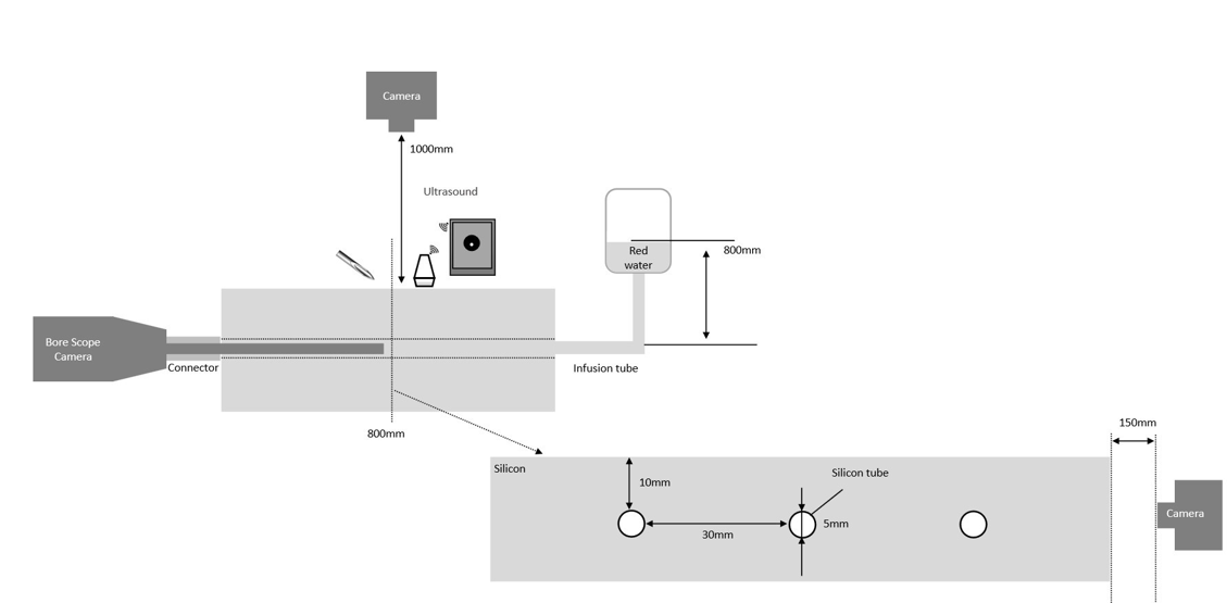
Figure1: Experimental setup for observation of the insertion process
The study of the first group of registered nurses from acute care hospitals in New York City who were not specialists in vascular access was conducted in May, 2025.
Participants were required to have worked at least two years in their current roles and have prior experience inserting USG GuidewireAssisted AIO midline catheters. The study sponsor was not disclosed. During the study, nurses reviewed blinded Instructions for Use (IFUs) for the Guidewire-Assisted AIO midline catheter, PowerGlide Pro™ (SKU: 4251628-02), selected because it is one of the most widely sold AIO midline products in the United States [23]. Each participant inserted the 3-step AIO midline catheter 5 times in accordance with the IFU.
The second group of vascular access specialists from acute care hospitals in New York City, who routinely perform USG Guidewire-Assisted AIO midline catheters, was conducted in September and November, 2025. Eligible participants were required to have a minimum of two years’ experience in their current roles. The study sponsor was not disclosed. During the study, participants reviewed blinded IFUs for two types of AIO midline catheters evaluated: Guidewire-Assisted PowerGlide Pro™ (3-step AIO midline catheter) and Non-Guidewire AIO midline SURFLO MIDELA™ (2-step AIO midline catheter), both currently available on the market. Each nurse inserted both catheter types 5 times each.
Data Collection and Analysis
Recorded videos of the procedures performed by participants under USG were reviewed and assessed to determine whether an extravascular or intravascular event occurred according to the predefined “event criteria” during any of the two or three steps in the insertion process (3-step Guidewire Assisted and 2-step Non-Guidewire AIO midline). An event was considered to have occurred if the needle tip and catheter deployment was performed extravascular, and if the needle reached the posterior wall or a needle had entered through the anterior wall and was retracted back out of the anterior wall, resulting in the catheter placement outside of the vessel. No event was considered to have occurred if the catheter was completely placed in the vessel without any occurrences consistent with the predefined event criteria. Furthermore, the final position of the catheter was assessed to determine whether it was placed in the vessel or out of vessel. In addition, the cumulative number of events occurring across the placement attempts and time to complete the steps were further assessed to determine during which step of the procedure they occurred.
Given the exploratory nature of this study, statistical hypothesis testing was not performed. Results were evaluated descriptively to identify potential patterns and trends Figure 2.
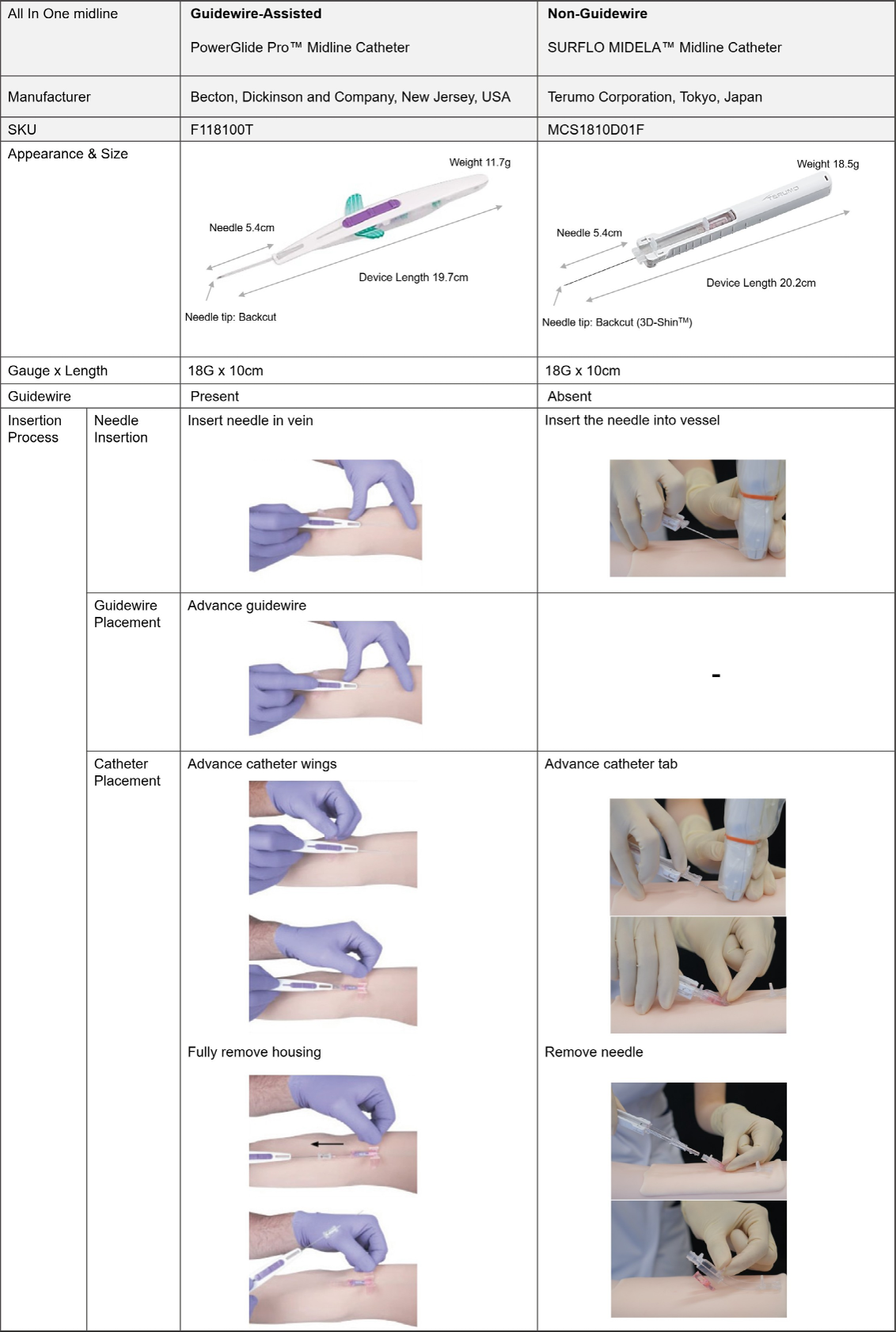
Figure 2: Comparing two types of AIO midlines during the insertion steps.
Results
Group 1: Identifying the steps during which adverse events occurred
A total of eight nurses participated in the study as shown in Table1 below. During screening, all but one reported being very familiar with USGPIV Table 1.
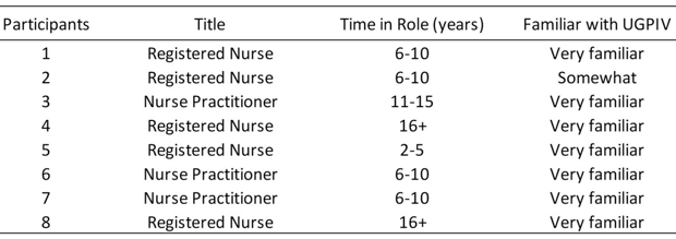
Table 1 Profile of Group1 participants.
The Figure 3 below shows that the catheter was placed outside the vein in 35% of insertions and in the vein in 65%. In total, 28 events occurred. Events occurred both when the catheter was placed in the vein and when the catheter was outside the vein. No event occurred in 12 insertions.
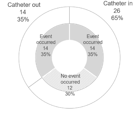
Figure 3: Catheter final position and events occurred.
As shown in the Figure 4 below, when examining each step in which these events occurred, 27 were observed during needle insertion under USG while visualizing the needle tip. 15 events occurred during guidewire placement, and 1 event occurred during catheter placement, when the needle slipped out of the vessel.
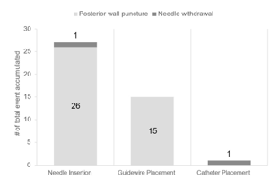
Figure 4: The cumulative number of events occurring across insertion steps
Figure 5 presents the box and whisker plots illustrating the time required for each procedural step. For the needle insertion step, the 25th percentile was 11 seconds, the median was 21 seconds, and the 75th percentile was 35 seconds. In contrast, the interquartile range (the middle 50% of the data) for the subsequent steps—Guidewire Placement and Catheter Placement-was shorter than that of the needle insertion step.
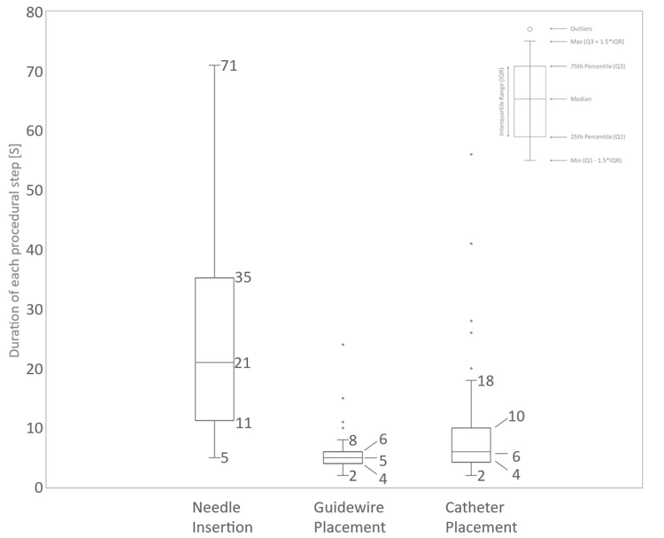
Figure 5: The duration of each step.
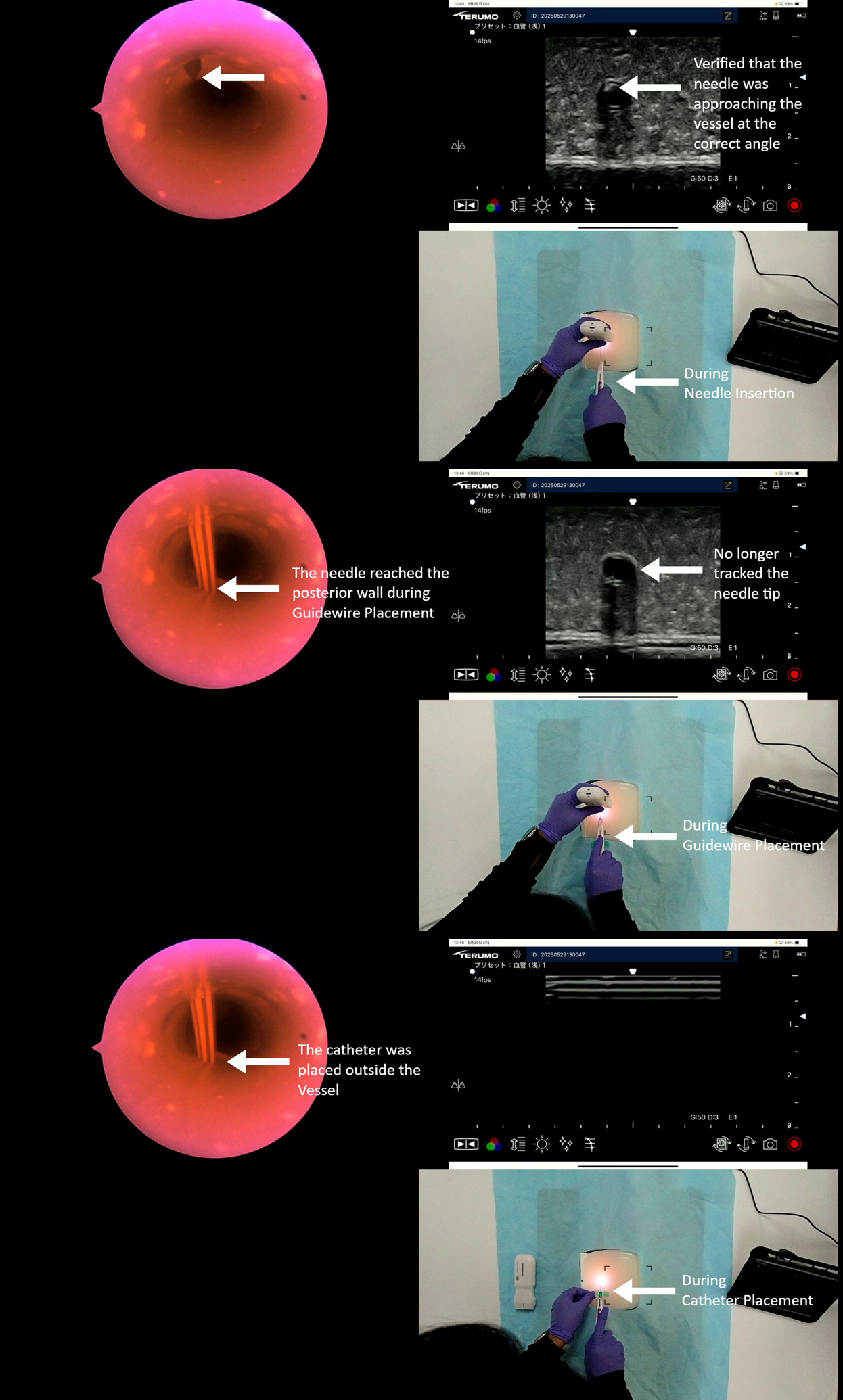
Figure 6: Representative of event occurred in the process of guidewire advancement and placed outside of the vessel.
A total of twelve nurses participated in the study as shown in Table 2. During screening, all reported being very familiar with UGPIV.
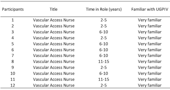
Table 2: Profile of Group 2 participants
As shown in Table 3 below, an analysis of each step in which these events occurred revealed that during needle insertion under USG while visualizing the needle tip, 6 events were observed in the Non-Guidewire group and 13 in the Guidewire-Assisted group. In the Guidewire-Assisted group, 5 events occurred during guidewire placement, whereas none were observed in the Non-Guidewire group. Furthermore, one event occurred during catheter placement in the Guidewire-Assisted group, while no such events were noted in the Non-Guidewire group.
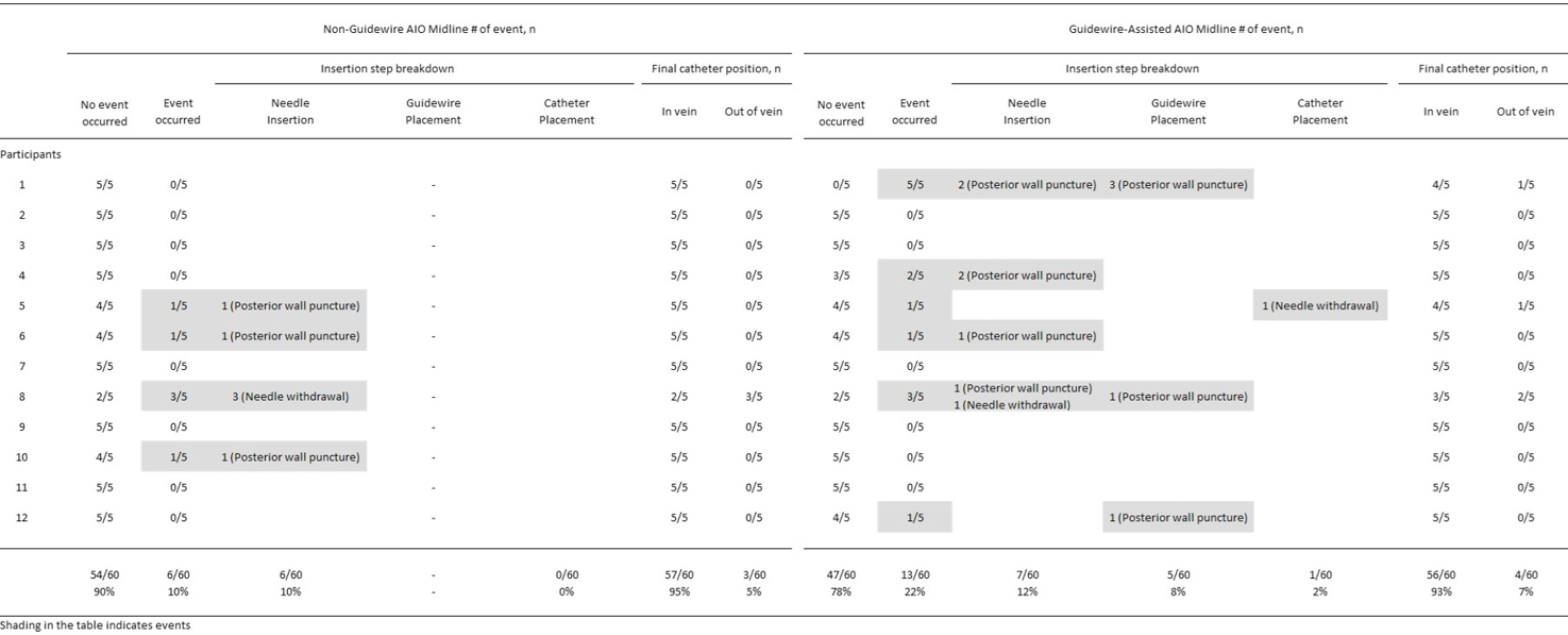
Table 3: Guidewire-Assisted vs Non-Guidewire.
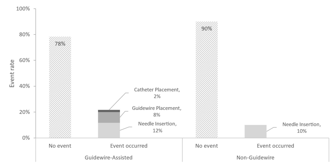
Figure 7: Result of Guidewire-Assisted vs Non-Guidewire
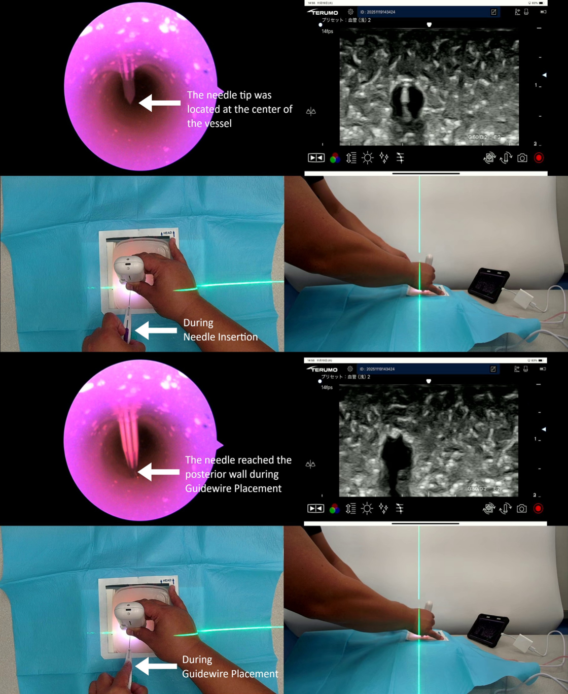
Figure 8: Representative of event occurred in the process of guidewire advancement.
Discussion
The three critical components for successful intravenous catheter placement are ultrasound assessment and needle tracking proficiency, hand stabilization and movement during catheter advancement, and manufacturer device ingenuity.
Ultrasound manufacturers have improved image clarity and reduction in real time image delay to assist clinicians with needle tracking. Vascular access device placement variation can occur due to clinician ultrasound interpretation. The user may identify the needle in an extravascular location but interpret it as intravascular or the user may identify the needle in the posterior wall of the vessel but interpret it as intravascular.
Vascular access device placement variation can occur due to hand stabilization even following successful USG needle placement into the vein. When a catheter is placed outside the vein it can typically go unnoticed until blood return is checked prior to infusion, potentially delaying treatment [3-4]. In this scenario the needle is properly placed within the vessel and confirmed, but as the ultrasound probe is placed down, and the hand moves to deploy the guidewire or catheter, the needle can be retracted outside of the vessel wall resulting in a proximal extravascular catheter placement or the needle pushed forward creating a posterior wall puncture extravascular placement. Posterior wall puncture is associated with hematoma formation and pain, which could also delay therapy. Furthermore, when posterior wall puncture
occurred and the catheter was still successfully placed, the risk of complications such as hematoma, infiltration, or extravasation may increase [3-4,27].
Midline catheters have evolved significantly in both design and insertion technique [13,21]. Early approaches utilized a modified Seldinger technique—separate needle access, followed by guidewire placement, then sheath introduction, and finally catheter advancement. This progressed to catheter-over-needle systems, which simplified the process but required advanced ultrasound skill to successfully “walk” the catheter through the vessel. More recently, innovations have led to catheter-over-needle devices with integrated guidewires. These designs enhance control during advancement, help reduce the risk of posterior vessel wall puncture, and improve overall success with catheter deployment.
The challenge with a Guidewire-Assisted device is that the tip of the needle may puncture the vessel and provide blood return, but until you “walk” the needle and catheter through the vessel wall the catheter will not be able to puncture the vessel wall despite the guidewire being thread. In addition, to deploy the guidewire requires hand movement that can affect catheter device stabilization and maintaining the needle in the intravascular space. It is in these transitions where the procedure variation is encountered.
In events in the first group (Figure 3), events occurred in 70% of cases among nurses who reported being “very familiar” with USG insertion, and in 35% of cases the catheter was ultimately placed outside the vein. USG insertion requires real time tracking of the needle tip, using the Probe-Needle technique (PN-T). Video analysis of the nurses’ tracking techniques demonstrated that almost all participants confirmed the vessel location and using the 3 stations of the needle approach, verified that the needle was approaching the vessel at the correct angle. However, once the needle entered the vessel, nurse participants no longer tracked the needle tip. The operators relied on tactile sensation after the needle entered the vessel and continued the subsequent steps accordingly. As a result, they did not recognize when the needle perforated the posterior wall or slipped out of the vessel, and advanced the catheter regardless, ultimately placing it extravascularly. Furthermore, because the needle tip was not visualized under ultrasound even after it had entered the vessel, events such as posterior wall puncture or needle withdrawal occurred repeatedly during all steps in the 3-step Guidewire-assisted processes—needle insertion, guidewire advancement, and catheter placement (Figure 4).
Although events still occurred among nurses proficient in USG insertion in Group 2, their frequency was markedly lower than Group1. When the insertion process was analyzed step by step, Guidewire-Assisted had 7 events and Non-Guidewire had 6 events during the needle insertion step in which the needle tip was visualized under ultrasound, yet, still punctured the posterior wall or exited the vein.
Although needle insertion was performed under ultrasound guidance to confirm intravascular needle tip position, maintaining this confirmation is not possible when the ultrasound probe is removed and the clinicians’ second hand is moved to the device to either deploy the catheter or deploy the guidewire. It was found that during this “maneuver” the needle tip can deviate leading to procedural failure. In the Guidewire-Assisted maneuver, the operator must place the ultrasound probe down and slide the guidewire forward which can inadvertently place a forward motion on the needle leading to the puncture of the posterior wall of the vessel, or a backward motion on the device leading to the needle migrating out of the vein. Finally, with the Guidewire-Assisted device, if the clinician is not using the PN-T and the catheter is not advanced beyond the vein wall, the needle can cannulate, the guidewire can be threaded, but the catheter not pass through the vein wall.
In contrast, the Non-Guidewire AIO catheter over the needle technique requires only one hand movement from the ultrasound probe to the catheter for deployment. This forward motion can cause the needle to puncture the back wall of the vessel, and the hand movement can cause the needle to migrate outside of the vessel prior to deployment. Yet, this forward or backward motion may be avoided by using the PN-T which allows the catheter to be walked in through the vein wall significantly prior to catheter deployment.
The needle insertion step may represent an important component of ultrasound-guided catheter placement, as variability observed during this phase appeared to be more operator dependent than subsequent procedural steps. In this exploratory study, devices with fewer procedural steps appeared to be associated with shorter procedure times and fewer observed variation among experienced ultrasound users. These findings are hypothesis-generating only and may suggest that Non-Guidewire AIO midline catheters may have the potential to support first-stick success.
Limitations
These exploratory findings from in-vitro investigation should be considered hypothesis-generating only and may warrant further investigation in appropriately powered studies.
Conclusions
Continuous ultrasound needle tip tracking is essential for safe midline insertion. The exploratory study shows that unrecognized tip deviation and posterior wall puncture drive placement failure and downstream complications. While AIO midline catheters resemble the traditional over the needle catheter placement, Guidewire-
Assisted devices require an additional step which may contribute to micro movements during insertion. These observations may suggest that device design could influence procedural performance. However, the findings should be considered hypothesis-generating and warrant further investigation.
Ethical Statement
This study was an observational study not conducted on patients. Informed consent for participation from the participants was obtained.
Conflicts of Interest
The authors declared the following potential conflicts of interest with respect to the research, authorship, and/or publication of this article: Matthew Ostroff providing research consultation for the Terumo Medical Corporation. Terumo funded the editorial assistance provided by Lois Kaufman and Catherine Adams and the research conducted by American Opinion Research.
The authors disclosed receipt of the following financial support for the research, authorship, and/or publication of this article: Matthew Ostroff provided manuscript support funded by Terumo Medical Corporation. American Opinion Research provided manuscript support and simulation research funded by Terumo Medical Corporation.
References
- Adžemović T, Govindan S, Zheutlin A, Horowitz J, Heath M, Kuhn L, et al. (2024) Awareness of peripheral IV catheters among nurses, physicians, and trainees: A prospective cohort study. Am J Infect Control 52: 1122-1127.
- Midline Catheters Show Fastest Growth Rate in Vascular Access A Global Market Analysis, Hadi Salempoor and Kamran Zamanian, Ph.D., iData Research Inc.
- Moureau N, Kaufman L (2025) Comparative study of peripheral intravenous catheter insertions with capillary, notched, and a grooved needle flashback design. https://doi.org/10.1177/11297298241313414 Volume 27, Issue 1.
- https://www.myamericannurse.com/wp-content/uploads/2019/09/ ant9-CE-Periperal-IV-821a.pdf, 2026.
- Van Loon FH, Buise MP, Claassen JJ, et al. (2018) Comparison of ultrasound guidance with palpation and direct visualization for peripheral vein cannulation in adult patients; a systematic review and meta-analysis. Br J Anaesth. 121: 358–366.
- Edwards C, Jones J (2018) Development and implementation of an ultrasound-guided peripheral intravenous catheter program for emergency nurses. J Emerg Nurs 44: 33–36.
- Costantino TG, Parikh AK, Satz WA, Fojtik JP (2005) Ultrasonographyguided peripheral intravenous access versus traditional approaches in patients with difficult intravenous access. Ann Emerg Med. 46: 456– 461.
- Bahl A, Pandurangadu AV, Tucker J, Bagan M (2016) A randomized IV placement in difficult access ED patients. Am J Emerg Med. 34: 1950–1954.
- Jørgensen R, Laursen CB, Konge L, Pietersen PI, Scand J (2021) Education in the placement of ultrasound-guided peripheral venous catheters: a systematic review, Trauma Resusc Emerg Med. 29.
- Salempoor S, Zamanian K, Ph.D., (2023) Midline Catheters Show Fastest Growth Rate In Vascular Access: A Global Market Analysis (2023) iData Research Inc., https://www.meddeviceonline.com/doc/ midline-catheters-show-fastest-growth-rate-in-vascular-access-aglobal-market-analysis-0001.
- Lee SW, (2022) Midline catheter insertion: focused on the procedure, 7(1): 27-30. DOI: https://doi.org/10.24184/tip.2022.7.1.27.
- Elli S, Pittiruti M, Pigozzo V, Cannizzo L, Giannini L, Siligato A, et al. (2020) Ultrasound-guided tip location of midline catheters.,Vasc 21:764-768.
- Dawson RB, Moureau NL, (2013) Midline Catheters: An Essential Tool in CLABSI Reduction Articles.
- Mun Hee Jeon, Cheol Seung Kim, Kyu Dam Han, Mi Jin Kim. (2022) Efficacy and Safety of Midline Catheters with Integrated Wire Accelerated Seldinger Technique.
- McKinney A DNP, CPNP-AC; Steanson K, et al. (2023) A Standardized Training Program in Ultrasound-Guided Intravenous Line Placement. Improving Nurses’ Confidence and Success.
- J. Gorgone M, O’Connor TP, (2022) Maximous SI How I Teach: Ultrasound-guided Peripheral Venous;3(4):598–609. doi: 10.34197/ ats-scholar.2022-0029HT
- Bruant A, Normand L (2025) Recent Advances in Ultrasound-Guided Peripheral Intravenous Catheter, Insertion. Nurs Rep. 15: 359.
- McMahon K, Thomas A, Isenberg JD (2025) Ultrasound-Guided Intravenous Access. PMID: 30252244 Bookshelf ID: NBK525988 In: Stat Pearls Treasure Island (FL): Stat Pearls Publishing.
- Jørgensen R, Laursen CB, Konge L, Pietersen PI (2021) Education in the placement of ultrasound-guided peripheral venous catheters: a systematic review. Scand J Trauma Resusc Emerg Med. 29: 83.
- Lee SW (2022) Midline catheter insertion: focused on the procedure. Trauma Image Proced 7: 27-30.
- https://www.bd.com/en-ca/products-and-solutions/products/productfamilies/powerglide-pro-midline-catheter, 2026.
- Mittelman A, MD. Common Pitfalls of Ultrasound-Guided Peripheral IV Placement 2020. https://www.emra.org/emresident/article/ultrasoundiv-placement-pitfalls.
- https://idataresearch.com/product/midline-market-size-share-andtrends-analysis-global-2024-2030-medcore/outcomes, 2023;102 e33624.
- https://www.accessdata.fda.gov/scripts/cdrh/cfdocs/cfmaude/search. cfm, 2026.
- Yamagami Y, Tomita K, Tsujimoto T, et al. (2017) Tourniquet application after local forearm warming to improve venodilation for peripheral intravenous cannulation in young and middle-aged adults: A single- blind prospective randomized controlled trial. Int J Nurs Stud. 72: 1-7.
- Helm RE, Klausner JD, Klemperer JD, Flint LM, Huang E (2015) Journal of Infusion Nursing. 38: 189-203.
- Andrea Dell’Amore, Chady Ammari, Rocco D’Andrea (2016) Peripheral venous catheter fracture with embolism into the pulmonary artery. PMCID: PMC5227288 PMID: 28149586.